


As biological analysis moves toward smaller samples, faster workflows, and increasingly complex diagnostic targets, microfluidics has become an important platform for integrating sample handling, biochemical reactions, enrichment, and signal detection. Yet the performance of a microfluidic system is closely tied to the precision of its physical architecture. Channel dimensions, fluidic interfaces, reaction chambers, valves, mixing structures, and detection regions can all influence how efficiently biological samples are processed.
By translating digital designs directly into complex three-dimensional microstructures, high-resolution 3D printing can help researchers develop customized microfluidic components that are difficult or time-consuming to manufacture using conventional fabrication methods. Three recent research studies illustrate this potential from different perspectives.
Case Study 1: Precision Microfluidics for Tumor Biomarker Analysis
Full paper → https://www.science.org/doi/10.1126/sciadv.aea2586
In a study published in Science Advances, researchers developed a dual-mode microfluidic immunostaining platform designed to address a practical challenge in rare tumor analysis: obtaining more diagnostic and biological information from limited tissue samples.
The platform combines different immunostaining workflows within a single microfluidic architecture, enabling diagnostic biomarker analysis alongside investigation of the tumor microenvironment. According to the study, the system can support analysis using only one to two tissue sections, substantially reducing tissue consumption while expanding the amount of information that can be obtained.
BMF's micro 3D printing played a key role in fabricating the physical components required for the fluidic system. Using microArch® S240 system (Optical resolution:10μm), researchers produced a 3D-printed fluid exchange manifold and molds for the microfluidic chip. The printed molds were subsequently used for PDMS replication, creating the microfluidic structures required for automated staining. The reported channel dimensional variation was below 1%, demonstrating the importance of high-resolution manufacturing for consistent microfluidic performance.
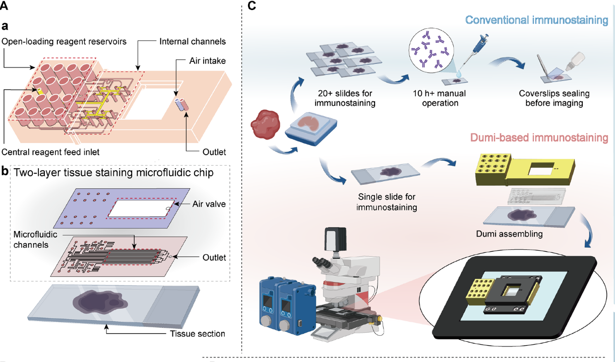
Fig. 3D-printed fluid exchange manifold and molds for the microfluidic chip.
For biomedical researchers, this can shorten the iteration cycle between device design and experimental validation, particularly when developing customized microfluidic architectures for specialized diagnostic workflows.
Case Study 2: Microfluidic Bacterial Detection with Integrated Signal Enhancement
Full paper → https://doi.org/10.1021/acs.analchem.5c02447
A second study explored microfluidics as a platform for rapid bacterial detection. Published in Analytical Chemistry, the research combined fluorogenic DNAzyme-based recognition with a photonic-crystal-assisted microfluidic architecture. In this study, researchers used BMF's 3D printing technology to construct μchip templates and prepared microfluidic structures using polydimethylsiloxane (PDMS) casting and encapsulation.
Rather than relying on a single detection mechanism, the platform integrates several functional stages, including a serpentine reaction region, magnetic-bead-based enrichment, and a photonic crystal module for optical signal enhancement. This architecture allows biological recognition and signal generation to be connected through controlled microscale fluid transport.
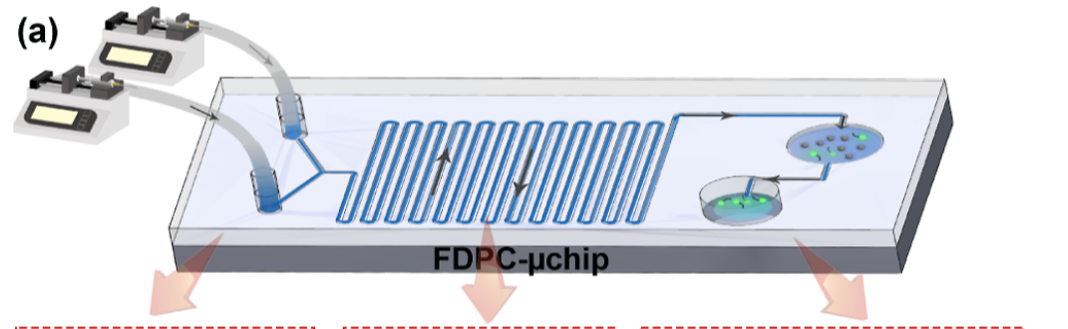
Fig. Working Principle of the FDPC-μchip
High-resolution 3D printing can provide researchers with a flexible route for prototyping customized microfluidic structures, helping them explore different channel configurations and functional layouts as diagnostic concepts evolve.
Case Study 3: Miniaturizing Sample Preparation for Cellular Trace Element Analysis
Full paper:https://doi.org/10.1021/acs.analchem.4c05810
Inductively coupled plasma mass spectrometry (ICP-MS) offers exceptional sensitivity for multi-element and isotope analysis, making it a powerful tool for investigating trace elements in biological systems. However, direct analysis of cellular samples can be challenging because conventional workflows often require large numbers of cells, while complex cellular matrices and extremely low analyte concentrations can compromise analytical performance.
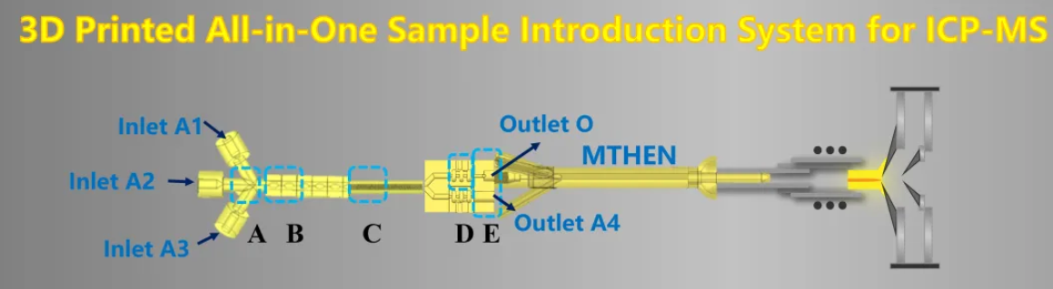
Fig. 3D printed ICP-MS
To address these limitations, by fabricating the integrated microextraction chip and customized nebulizer with the BMF microArch® S130 (Optical resolution: 2 μm), the research team developed a compact sample introduction platform capable of integrating cell lysis, microextraction, fluidic control, and nebulization for online ICP-MS analysis. The system also incorporated a 3D-printed microflow high-efficiency nebulizer (3DP-MTHEN), whose customized geometry and interface enabled direct integration with the ICP-MS torch. This demonstrated how high-resolution additive manufacturing can extend beyond individual microfluidic chips to the fabrication of customized analytical interfaces.
What Micro 3D Printing Adds to Microfluidic Bioanalysis
Across these three research directions, the applications are different, but the underlying manufacturing requirement is similar: biological analysis increasingly depends on precisely engineered microscale structures. BMF's PμSL technology is designed around this need for high-resolution and customized manufacturing. With systems spanning different precision levels, BMF provides researchers with a platform for developing microfluidic structures across a range of application requirements.
As microfluidic diagnostics continue to pursue smaller sample volumes, higher integration, faster analysis, and more personalized testing, high-resolution 3D printing can provide researchers with greater freedom to design and iterate the physical structures behind these systems.
For researchers developing next-generation lab-on-a-chip, biosensing, cellular analysis, and diagnostic platforms, micro 3D printing is therefore becoming more than a prototyping technology. It is increasingly serving as an enabling manufacturing tool for translating complex microfluidic concepts into functional analytical systems.