


Liposomes are widely investigated as nanoscale carriers for drug delivery, molecular imaging, diagnostics, and targeted therapy. Their ability to encapsulate therapeutic or imaging agents within a lipid bilayer makes them attractive for applications where controlled transport and biological targeting are required. However, the performance of a liposomal system is closely linked to how precisely its preparation environment can regulate fluid mixing, mass transfer, reaction conditions, and surface functionalization.
Microfluidic technology has emerged as an important platform for addressing these challenges. By controlling multiple liquid phases within microscale channels, microfluidic devices can provide more predictable hydrodynamic conditions than many conventional bulk preparation methods.
This is where high-precision micro 3D printing provides an important manufacturing advantage. BMF's Projection Micro Stereolithography (PμSL) technology enables the rapid fabrication of complex microfluidic structures with optical resolutions down to 2μm, creating new possibilities for researchers developing customized platforms for liposome synthesis, functionalization, analysis, and biomedical testing.
Case Study 1: Microfluidics for Automated Manipulation of Extracellular Vesicles
Full paper → https://doi.org/10.1021/acsnano.5c14524
Exosomes are emerging as promising biomarkers for liquid biopsy because their membrane proteins can provide valuable information about disease states. However, their nanoscale dimensions, heterogeneous composition, and complex biological background make efficient separation and target-specific aptamer screening challenging.
A research team developed DREAMbot, an automated robotic platform integrating microfluidics for iterative exosome-targeted aptamer screening. A key component of DREAMbot is its deterministic lateral displacement (DLD) sorting chip, which enables size-based separation of exosomes from larger cellular components and extracellular vesicles.
To fabricate the microscale pillar-array structures required for precise DLD separation, the research team used BMF's microArch® S240, based on Projection Micro Stereolithography (PμSL) technology, with 10 μm optical resolution. PμSL enables the rapid fabrication of intricate three-dimensional microstructures with high dimensional fidelity. This capability was particularly valuable for producing the micro-pillar array architecture and dual-region separation structures required by the DLD chip.
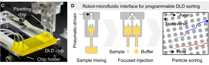
Fig1.Using BMF's PμSL technology to fabricate the DLD chip
The resulting chip achieved a reported exosome recovery rate above 90%, providing an efficient front-end separation step for the subsequent aptamer screening workflow. By integrating precision-fabricated microfluidic components with robotic automation, DREAMbot reduces the dependence on repetitive manual operations and improves consistency across multiple rounds of selection.
Further testing with clinical serum samples demonstrated substantially stronger binding signals for exosomes derived from liver cancer patients than for healthy controls, highlighting the potential of the resulting aptamers for liquid-biopsy applications.
Case Study2 : BMF PμSL 3D Printing Enables a Biomimetic Micromixer for Machine Learning-Guided Liposome Manufacturing
Full paper → https://doi.org/10.26599/NR.2025.94907713
A second case provides a more direct example of microfluidic liposome engineering. Chen and colleagues developed a specialized micromixer for the one-step production of targeted emodin liposomes. The reported platform uses a combination of biomimetic groove structures and splitting-and-recombination channels to enhance mixing between different liquid streams. Machine learning was subsequently introduced to help predict liposome particle size from process parameters.
Liposomes are promising drug delivery vehicles for improving the solubility, stability, and targeted delivery of therapeutic compounds. However, conventional liposome fabrication methods often involve multiple processing steps and can suffer from limited batch-to-batch consistency. Microfluidic manufacturing offers a more controllable alternative, but efficient mixing within microscale channels remains a critical challenge for producing liposomes with precisely controlled properties.
To address this challenge, a research team developed a biomimetic leaf-vein groove–horseshoe micromixer (VGHM) and combined it with machine learning to enable controlled, one-step fabrication of targeted emodin-loaded liposomes for ulcerative colitis therapy. The researchers used BMF's Projection Micro Stereolithography (PμSL) technology with 25 μm optical resolution, to fabricate the master mold. The 3D-printed mold was subsequently replicated using PDMS molding and plasma bonding, producing the final microfluidic device with the designed channel geometry and enclosed flow path.
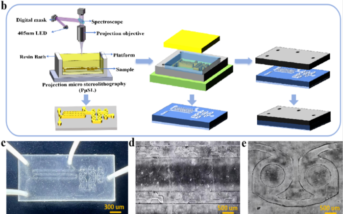
Fig2. Preparation process and printing mold
This fabrication route allowed the researchers to rapidly realize the customized three-dimensional microchannel architecture required by the VGHM design, demonstrating the value of high-precision 3D printing as an enabling technology for rapid prototyping and functional development of microfluidic devices.
Case Study 3: 3D-Printed Microfluidics for Aptamer-Modified Liposomal Probes
Full paper→https://doi.org/10.1016/j.snb.2024.136538
Targeted contrast agents play an important role in improving the specificity and sensitivity of biomedical imaging. Liposomes can encapsulate imaging agents while providing a versatile platform for surface functionalization with targeting molecules such as aptamers. However, conventional liposome fabrication methods can involve multiple processing steps and may produce variations in particle size and distribution, making consistent preparation of functionalized nanoprobes challenging.
BMF's microArch® S140 (Optical resolution: 10μm) system was used to fabricate the microfluidic chip, which incorporated a dedicated mixing channel and micropillar-based mixing region, allowing the researchers to manipulate fluid flow and enhance mixing between the lipid-containing organic phase and aqueous phase.
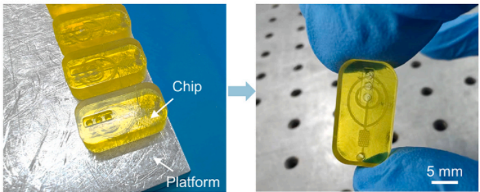
Fig3. 3D-printed microfluidic mixer
Using the 3D-printed microfluidic mixer, the team developed a rapid preparation strategy for methylene blue-loaded, anti-PD-L1 aptamer-functionalized liposomes (Apt-MB-Lip). The microfluidic approach provided controlled fluid interaction at the microscale, supporting the formation of relatively uniform liposomes while integrating aptamer modification into the nanoprobe preparation workflow.
The researchers subsequently characterized the resulting liposomes through particle-size analysis, PDI, zeta-potential measurements, TEM imaging, and UV-Vis spectroscopy. These analyses confirmed the formation of the liposomal structures, methylene blue encapsulation, and successful aptamer functionalization.
BMF: Enabling the Next Generation of Microfluidic Biofabrication
The future of microfluidic liposome technology is unlikely to be limited to particle formation alone. Increasingly, researchers are combining nanocarrier synthesis with targeting, purification, biological analysis, imaging, and drug screening.
The three research directions considered here illustrate this evolution from different perspectives. Automated microfluidics can improve the handling and selection of nanoscale biological vesicles; engineered micromixers can provide greater control over liposome formation and functionalization; and precision 3D-printed microfluidic chips can translate these principles into customized biomedical platforms.
BMF's PμSL technology provides researchers with a direct pathway from digital microfluidic design to physical experimentation. With systems supporting resolutions down to 2 μm and dedicated applications in microfluidics and biomedicine, BMF enables the development of complex microstructures for emerging life-science applications.
From engineered micromixers and liposome synthesis chips to integrated vesicle-processing and biomedical analysis platforms, high-precision micro 3D printing can help shorten the distance between an innovative microfluidic concept and a functional prototype.